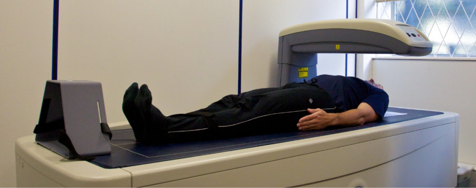
Bone Densitometry (DEXA, DXA)
May 20, 2019
Bone densitometry, also called dual-energy x-ray absorptiometry, DEXA or DXA,
uses a very small dose of ionizing radiation to produce pictures of the inside of the
body (usually the lower (or lumbar) spine and hips) to measure bone loss. It is
commonly used to diagnose osteoporosis, to assess an individual’s risk for
developing osteoporotic fractures. DXA is simple, quick and noninvasive. It’s also
the most commonly used and the most standard method for diagnosing
osteoporosis.
Bone density testing is strongly recommended if you:
- are a post-menopausal woman and not taking estrogen.
- have a personal or maternal history of hip fracture or smoking.
- are a post-menopausal woman who is tall (over 5 feet 7 inches) or thin (less than 125 pounds).
- are a man with clinical conditions associated with bone loss, such as rheumatoid arthritis, chronic kidney or liver disease.
- use medications that are known to cause bone loss, including corticosteroids such as Prednisone, various anti-seizure medications such as Dilantin and certain barbiturates, or high-dose thyroid replacement drugs.
- have type 1 (formerly called juvenile or insulin-dependent) diabetes, liver disease, kidney disease or a family history of osteoporosis.
- have high bone turnover, which shows up in the form of excessive collagen in urine samples.
- have a thyroid condition, such as hyperthyroidism.
- have a parathyroid condition, such as hyperparathyroidism.
- have experienced a fracture after only mild trauma.
- have had x-ray evidence of vertebral fracture or other signs of osteoporosis.
The Vertebral Fracture Assessment (VFA), a low-dose x-ray examination of the
spine to screen for vertebral fractures that is performed on the DXA machine, may be recommended for older patients, especially if:
they have lost more than an inch of height.
- have unexplained back pain.
- if a DXA scan gives borderline readings.
- the DXA images of the spine suggest a vertebral deformity or fracture.
More s


