Radiology
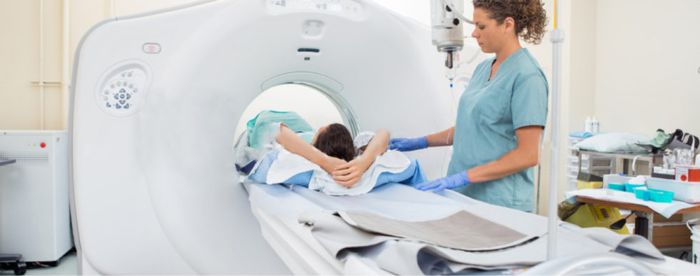
CT scan
A computerised tomography (CT) scan uses X-rays and a computer to create detailed images of the inside of the body When CT scans are used
CT scans can produce detailed images of many structures inside the body, including the internal organs, blood vessels and bones. They can be used to:
- diagnose conditions – including damage to bones, injuries to internal organs, problems with blood flow, stroke, and cancer
- guide further tests or treatments – for example, CT scans can help determine the location, size and shape of a tumour before having radiotherapy, or allow a doctor to take a needle biopsy (where a small tissue sample is removed using a needle) or drain an abscess
- monitor conditions – including checking the size of tumours during and after cancer treatment
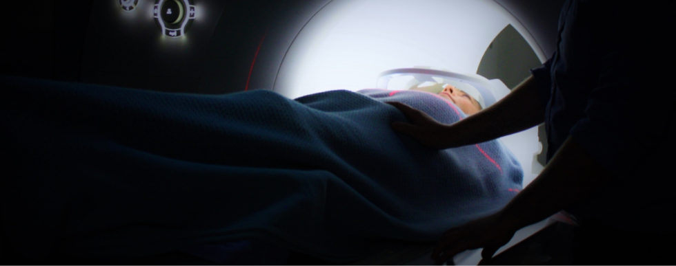
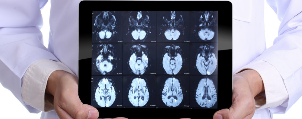
MRI
Magnetic resonance imaging (MRI) is a type of scan that uses strong magnetic fields and radio waves to produce detailed images of the inside of the body. An MRI scan can be used to examine almost any part of the body, including the:
- brain and spinal cord
- bones and joints
- breasts
- heart and blood vessels
- internal organs, such as the liver, womb or prostate gland
X-RAY
X-rays are a form of electromagnetic radiation that can pass through solid objects, including the body. X-rays penetrate different objects more or less according to their density. In medicine, X-rays are used to view images of the bones and other structures in the body. The most common form of X-ray used is X-ray radiography, which can be used to help detect or diagnose:
- Bone fractures
- Infections (such as pneumonia)
- Calcifications (like kidney stones or vascular calcifications)
- Some tumors
- Arthritis in joints
- Bone loss (such as osteoporosis)
- Dental issues
- Heart problems (such as congestive heart failure)
- Blood vessel blockages
- Digestive problems
- Foreign objects (such as items swallowed by children)
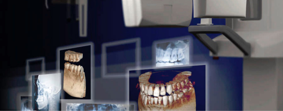
Panorama xray
Dental X-rays (radiographs) are images of your teeth that your dentist uses to evaluate your oral health. These X-rays are used with low levels of radiation to capture images of the interior of your teeth and gums. This can help your dentist to identify problems, like cavities, tooth decay, and impacted teeth.
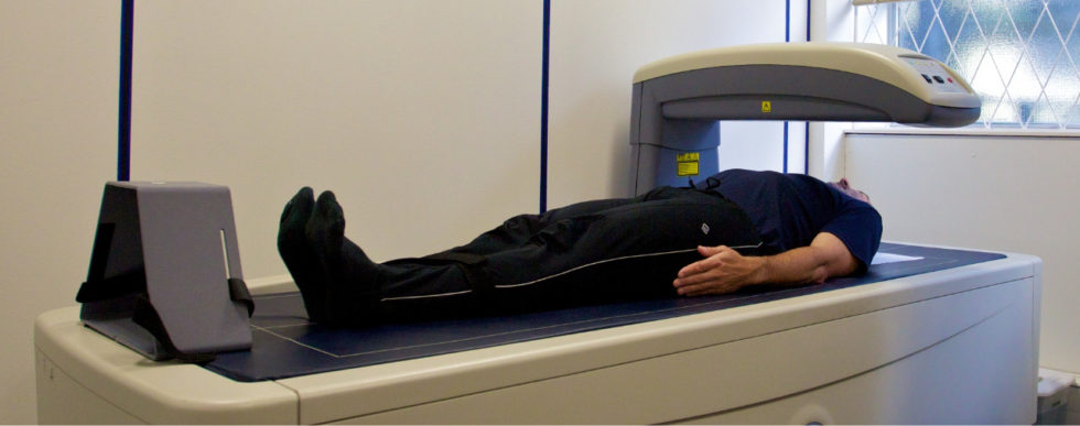
Bone Densitometry (DEXA, DXA)
Bone densitometry, also called dual-energy x-ray absorptiometry, DEXA or DXA, uses a very small dose of ionizing radiation to produce pictures of the inside of the body (usually the lower (or lumbar) spine and hips) to measure bone loss. It is commonly used to diagnose osteoporosis, to assess an individual's risk for developing osteoporotic fractures. DXA is simple, quick and noninvasive. It's also the most commonly used and the most standard method for diagnosing osteoporosis.
Bone density testing is strongly recommended if you:
- are a post-menopausal woman and not taking estrogen.
- have a personal or maternal history of hip fracture or smoking.
- are a post-menopausal woman who is tall (over 5 feet 7 inches) or thin (less than 125 pounds).
- are a man with clinical conditions associated with bone loss, such as rheumatoid arthritis, chronic kidney or liver disease.
- use medications that are known to cause bone loss, including corticosteroids such as Prednisone, various anti-seizure medications such as Dilantin and certain barbiturates, or high-dose thyroid replacement drugs.
- have type 1 (formerly called juvenile or insulin-dependent) diabetes, liver disease, kidney disease or a family history of osteoporosis.
- have high bone turnover, which shows up in the form of excessive collagen in urine samples.
- have a thyroid condition, such as hyperthyroidism.
- have a parathyroid condition, such as hyperparathyroidism.
- have experienced a fracture after only mild trauma.
- have had x-ray evidence of vertebral fracture or other signs of osteoporosis.
The Vertebral Fracture Assessment (VFA), a low-dose x-ray examination of the spine to screen for vertebral fractures that is performed on the DXA machine, may be recommended for older patients, especially if: they have lost more than an inch of height.
- have unexplained back pain.
- if a DXA scan gives borderline readings.
- the DXA images of the spine suggest a vertebral deformity or fracture.
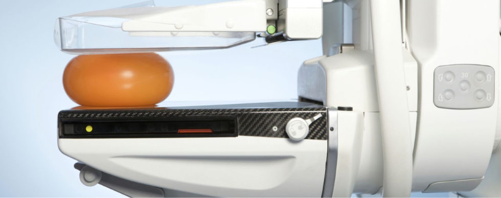
Mammography
Mammography is a specific type of breast imaging that uses low-dose x-rays to detect cancer early – before women experience symptoms – when it is most treatable. Tell your doctor about any breast symptoms or problems, prior surgeries, hormone use, whether you have a family or personal history of breast cancer, and if there's a possibility you are pregnant. If possible, obtain copies of your prior mammograms and make them available to your radiologist on the day of your exam. Leave jewelry at home and wear loose, comfortable clothing. You may be asked to wear a gown. Don't wear deodorant, talcum powder or lotion under your arms or on your breasts as these may appear on the mammogram and interfere with correct diagnosis.
Breast tomosynthesis, also called three-dimensional (3-D) mammography and digital breast tomosynthesis (DBT), is an advanced form of breast imaging where multiple images of the breast from different angles are captured and reconstructed ("synthesized") into a three-dimensional image set. In this way, 3-D breast imaging is similar to computed tomography (CT) imaging in which a series of thin "slices" are assembled together to create a 3-D reconstruction of the body. Although the radiation dose for some breast tomosynthesis systems is slightly higher than the dosage used in standard mammography, it remains within the FDA- approved safe levels for radiation from mammograms. Some systems have doses very similar to conventional mammography.
Large population studies have shown that screening with breast tomosynthesis results in improved breast cancer detection rates and fewer "call-backs," instances where women are called back from screening for additional testing because of a potentially abnormal finding.
Breast tomosynthesis may also result in:
- earlier detection of small breast cancers that may be hidden on a conventional mammogram
- greater accuracy in pinpointing the size, shape and location of breast abnormalities
- fewer unnecessary biopsies or additional tests
- greater likelihood of detecting multiple breast tumors
- clearer images of abnormalities within dense breast tissue

Ultrasound
Ultrasound imaging uses sound waves to produce pictures of the inside of the body. It is used to help diagnose the causes of pain, swelling and infection in the body's internal organs and to examine a baby in pregnant women and the brain and hips in infants. It's also used to help guide biopsies, diagnose heart conditions, and assess damage after a heart attack. Ultrasound is safe, noninvasive, and does not use ionizing radiation.
What are some common uses of the procedure? Ultrasound examinations can help to diagnose a variety of conditions and to assess organ damage following illness.
Ultrasound is used to help physicians evaluate symptoms such as:
- pain
- swelling
- infection
Ultrasound is a useful way of examining many of the body's internal organs, including but not limited to the:
- heart and blood vessels, including the abdominal aorta and its major branches
- liver
- gallbladder
- spleen
- pancreas
- kidneys
- bladder
- uterus, ovaries, and unborn child (fetus) in pregnant patients
- eyes
- thyroid and parathyroid glands
- scrotum (testicles)
- brain in infants
- hips in infants
- spine in infants
Ultrasound is also used to:
- guide procedures such as needle biopsies, in which needles are used to sample cells from an abnormal area for laboratory testing.
- image the breasts and guide biopsy of breast cancer.
- diagnose a variety of heart conditions, including valve problems and congestive heart failure, and to assess damage after a heart attack.
Ultrasound of the heart is commonly called an "echocardiogram" or "echo" for short.
Doppler ultrasound images can help the physician to see and evaluate:
- blockages to blood flow (such as clots)
- narrowing of vessels
- tumors and congenital vascular malformations
- reduced or absent blood flow to various organs
- greater than normal blood flow to different areas, which is sometimes seen in infections